



Have you ever been asked to get a dental X ray and wondered, “Is it really necessary?” or “Are dental X rays safe?” These are common questions, especially when the word radiation is involved. A dental X ray uses a small amount of radiation to create images of your teeth, roots, jawbone, and other areas that cannot always be seen during a regular dental examination.
Have you ever been asked to get a dental X ray and wondered, “Is it really necessary?” or “Are dental X rays safe?” These are common questions, especially when the word radiation is involved. A dental X ray uses a small amount of radiation to create images of your teeth, roots, jawbone, and other areas that cannot always be seen during a regular dental examination.
The good news is that modern dental X rays involve very low radiation exposure. Dentists also follow radiation safety practices to avoid unnecessary exposure. So, if you have been advised to get a dental X ray, understanding why it is needed and how it is performed can make the process much less worrying.
Quick Overview
Dental X rays are generally safe when they are clinically needed and performed using appropriate safety measures. They use a very small amount of radiation and help dentists detect problems that may not be visible during a regular examination. The type and frequency of dental X rays should depend on your age, oral health, symptoms, and individual risk factors.
What Are Dental X-Rays?
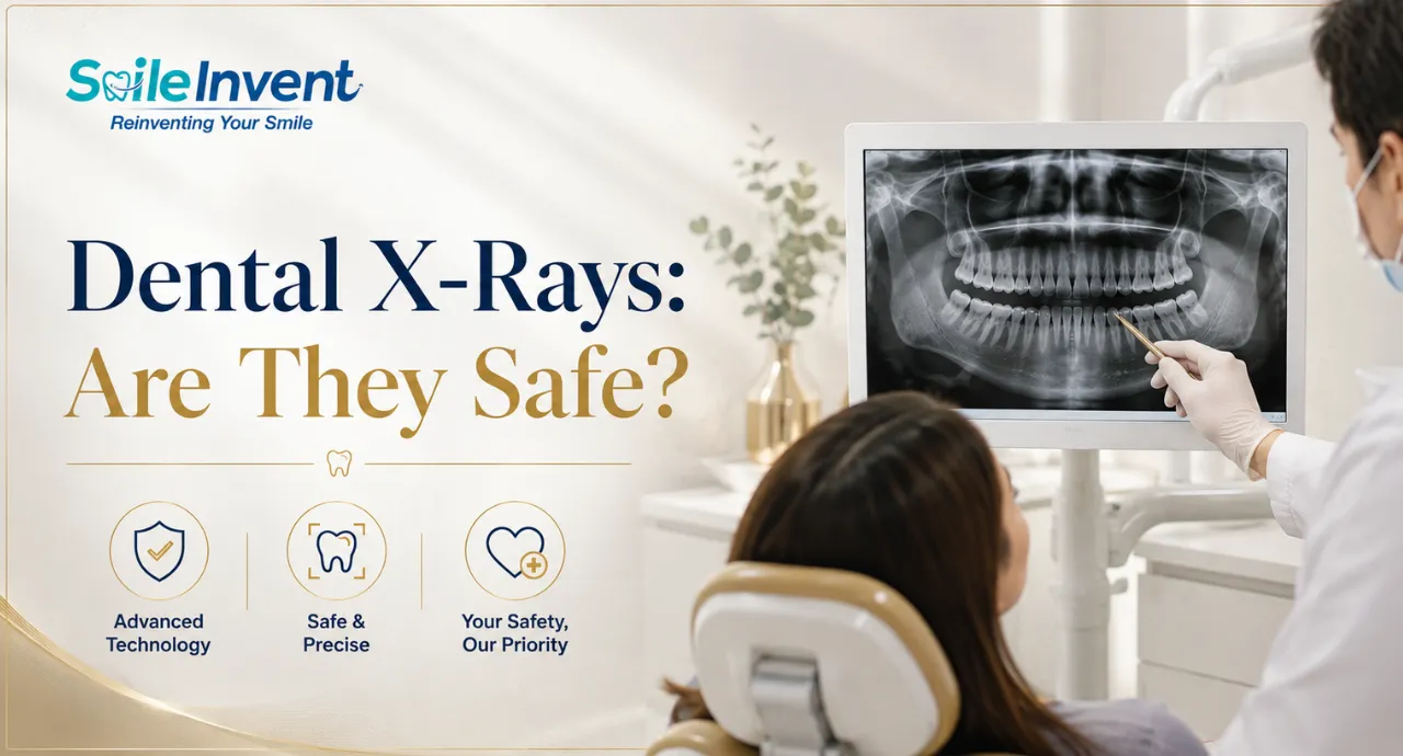
Dental X rays, also called dental radiographs, are images of the teeth and mouth created using X ray radiation. They allow dentists to see areas beneath the surface of the teeth and gums, including tooth roots, jawbone and other structures that cannot be examined properly just by looking inside the mouth.
During a dental X ray, a small amount of radiation passes through the area being examined. Different tissues absorb the radiation differently, which creates an image that the dentist can study. Modern dental practices commonly use digital imaging, where the image is captured using a digital sensor and can be viewed quickly on a computer.
This is where dental radiology becomes useful. A dentist can combine the X ray findings with your symptoms, medical and dental history, and physical examination to get a clearer picture of what is happening.
Why Do Dentists Recommend Dental X-Rays?
A normal dental examination can reveal many problems, but some conditions develop between teeth, beneath existing fillings, around tooth roots, or inside the jawbone. A dental X ray can provide information that is difficult or impossible to see during a visual examination.
Your dentist may recommend a tooth X ray to:
- Find cavities between teeth or beneath existing restorations.
- Check for infections or abscesses around tooth roots.
- Look for bone loss linked to gum disease.
- Identify impacted or unerupted teeth.
- Examine wisdom teeth and their position.
- Check for dental or jaw injuries.
- Plan treatments such as implants, braces, root canals or extractions.
- Monitor healing after certain dental procedures.
Dental X rays can sometimes identify cavities before they become visible during a regular examination. This makes them useful when your dentist needs a clearer view before deciding on treatment.
Types of Dental X-Rays
Not every dental X ray shows the same area. Your dentist chooses the type of image based on what they need to examine.
The choice depends on the information your dentist needs. A more advanced scan is not automatically better. Current guidance recommends using the lowest exposure option that can provide the information needed for diagnosis or treatment planning.
Are Dental X-Rays Safe?
Yes. Dental X rays are generally considered safe when they are clinically necessary and performed correctly. The amount of radiation used for routine dental imaging is very low. Still, X rays use ionizing radiation, so dentists avoid taking images that are not needed.
The latest ADA recommendations emphasize that dental X rays should be based on a patient's individual needs. Before recommending an image, the dentist should consider the patient's dental history, current oral health, symptoms, previous X rays and risk of dental disease. This means there is no single X ray schedule that is right for everyone.
The goal is simple: get the information needed for proper care while keeping radiation exposure as low as reasonably achievable. This approach is commonly referred to as the ALARA principle.
How Much Radiation Do Dental X-Rays Use?
The amount of radiation from a dental X ray is very small compared with many other forms of medical imaging. The exact exposure depends on the type of image, the equipment, the settings and the area being examined.
That does not mean radiation should be ignored. Repeated or unnecessary exposure should still be avoided. This is why dentists are encouraged to prescribe X rays only when they can provide useful information and to use appropriate dose reduction methods.
Digital imaging, proper positioning, limiting the X ray beam to the area being examined and using appropriate exposure settings are some of the ways dental professionals can reduce unnecessary radiation.
Digital Dental X-Rays vs Traditional X-Rays
Most modern dental clinics use digital imaging because it allows images to be captured, viewed and stored electronically. Traditional film X rays require photographic film and chemical processing.
Digital dental X rays can also make it easier for dentists to review images and compare them with previous records when needed.
Are Dental X-Rays Safe for Children?
Parents often have extra concerns about dental X rays because children are still growing. The answer is reassuring: dental X rays can be used safely in children when they are clinically necessary and the exposure is properly managed.
Children should not receive X rays simply because it is a routine part of every visit. The dentist should first examine the child and decide whether an image is needed. Factors such as age, stage of dental development, cavity risk, symptoms and previous dental images can influence that decision.
For example, an X ray may be useful when a dentist needs to check developing permanent teeth, look for cavities between baby or permanent teeth, assess an unerupted tooth or plan orthodontic treatment.
The ADA also supports child specific approaches to dental imaging, including using appropriate exposure settings and limiting the X ray beam to the area that needs to be examined.
Are Dental X-Rays Safe During Pregnancy?
If you are pregnant or think you may be pregnant, tell your dentist before having a dental X ray. This allows your dentist to consider your pregnancy along with your dental symptoms and treatment needs.
Current guidance from the American Academy of Oral and Maxillofacial Radiology states that dental X rays are safe during pregnancy when they are clinically needed. The radiation dose from dental imaging is extremely low, and appropriate imaging should not be delayed when it is necessary for diagnosis or treatment.
The key point is not to avoid all dental X rays during pregnancy. Instead, the dentist should decide whether the image is actually needed and use appropriate radiation safety practices.
What Happens During a Dental X-Ray?
A dental X ray is usually a quick procedure. You do not feel the X rays themselves, although some people may find holding the sensor inside the mouth slightly uncomfortable.
Here is what usually happens:
- The dental professional positions the sensor or imaging device.
- You may be asked to bite gently on a small holder for certain types of X rays.
- Your head or jaw is positioned so the correct area can be captured.
- You are asked to stay still while the image is taken.
- The image is checked and reviewed by the dentist.
How Often Should You Get Dental X-Rays?
There is no fixed answer to how often every person should get a dental X ray. You may need them more or less often depending on your oral health and risk of dental problems.
Your dentist may consider:
- Your age and stage of dental development.
- History of cavities.
- Gum health.
- Current symptoms such as pain or swelling.
- Previous dental treatment.
- Your risk of developing new dental problems.
- Previous X ray images that are already available.
For someone with healthy teeth and a low risk of dental disease, frequent imaging may not be necessary. Someone with symptoms, active dental disease or a higher risk of problems may need imaging sooner. The ADA's 2026 recommendations specifically support individualized decisions rather than a one size fits all schedule.
Can Dental X-Rays Detect Problems That Cannot Be Seen?
Yes. This is one of the main reasons dentists use dental X rays.
A tooth can look normal from the outside while a problem is developing underneath or between the teeth. A dental X ray can help reveal changes that may not yet be visible during a regular examination.
For example, X rays can help detect:
- Cavities between teeth.
- Decay beneath existing fillings or restorations.
- Infection around a tooth root.
- Bone loss related to gum disease.
- Impacted or unerupted teeth.
- Some abnormalities affecting the teeth or jaw.
- Fractures or injuries in certain areas.
MedlinePlus notes that dental X rays can reveal cavities before they are clinically visible. Cleveland Clinic also lists infections, bone loss and impacted teeth among conditions that dental X rays can help identify.
Dental X-Rays vs CBCT: What's the Difference?
Both are forms of dental imaging, but they are used for different purposes.
A conventional dental X ray generally produces a two dimensional image, while a CBCT scan creates a three dimensional view of the teeth, jaw and surrounding structures.
A CBCT scan is not automatically a better choice just because it provides more information. Current ADA guidance recommends using CBCT when the additional information is clinically necessary and lower exposure options cannot provide what the dentist needs.
How Dentists Keep Dental X-Ray Exposure Low
Dental X ray safety is not only about the machine. It also depends on when the image is prescribed and how the examination is performed.
Dentists can reduce unnecessary exposure by:
- Recommending X rays based on clinical need.
- Reviewing previous dental images when they are available.
- Choosing the appropriate type of X ray for the problem.
- Using digital imaging where appropriate.
- Limiting the X ray beam to the area being examined.
- Positioning the patient correctly to avoid repeat images.
- Using appropriate exposure settings.
- Choosing CBCT only when the required information cannot be obtained with a lower exposure option.
Important
Current ADA guidance has also changed older advice around routine lead aprons and thyroid collars during dental imaging. They are no longer routinely recommended for dental X ray procedures because proper beam restriction, positioning and dose control are more effective ways to minimize unnecessary exposure. Dental clinics should follow current professional guidance and applicable local regulations.
How Smile Invent Dental Clinic Uses Dental X-Rays for Better Diagnosis
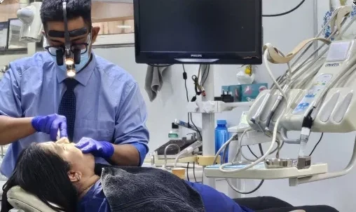
At Smile Invent Dental Clinic, a dental X ray is not something a patient needs simply because they have come for a checkup. The dentist first examines your teeth, gums, symptoms and dental history. If an X ray can provide information that cannot be obtained through a regular examination, the dentist may recommend it as part of the diagnostic process.
A tooth X ray can help the dentist understand what is happening beneath the visible surface. For example, if you have unexplained tooth pain, suspected decay, swelling, an impacted wisdom tooth or a concern around a previous dental treatment, an X ray may provide useful information for diagnosis and treatment planning.
The team at Smile Invent Dental Clinic can explain why an image is being recommended and what the dentist is looking for. This patient focused approach helps ensure that dental radiology is used as a diagnostic aid rather than as an unnecessary routine procedure. If you have been advised to get a dental X ray or are experiencing a dental problem that may require further investigation, a consultation can help determine what imaging, if any, is appropriate for you.
FAQ’s
1. Are dental X rays safe?
Yes. Dental X rays are generally safe when they are clinically necessary and performed using appropriate radiation safety measures. The radiation exposure from routine dental imaging is very low.
2. Are dental X rays safe for children?
Yes. Children can safely have dental X rays when they are needed for diagnosis or treatment planning. Dentists should consider the child's age, dental development, risk factors and symptoms before recommending imaging.
3. Are dental X rays safe during pregnancy?
Yes, when clinically needed. Tell your dentist if you are pregnant or may be pregnant so they can consider your situation before recommending imaging. The AAOMR states that dental X rays can be safely performed during pregnancy.
4. Do dental X rays hurt?
No. The X ray itself does not cause pain. Some people may find holding the sensor in the mouth uncomfortable or may experience a gag reflex during certain types of dental imaging.
5. Can a dental X ray detect cavities?
Yes. Dental X rays can help dentists find cavities, including decay between teeth that may not be visible during a regular dental examination.
6. How often should I get dental X rays?
There is no fixed schedule that applies to everyone. Your dentist decides when imaging is needed based on your oral health, age, symptoms, cavity risk, previous dental images and treatment needs.
7. What is the difference between a dental X ray and a CBCT scan?
A conventional dental X ray generally provides a 2D image, while CBCT produces a 3D image. CBCT can provide more detailed information for selected complex cases, but it should be used only when the additional information is clinically necessary.
8. Can I refuse a dental X ray?
You can discuss your concerns and options with your dentist. If an X ray is recommended, ask what the dentist is looking for and whether previous images or another diagnostic approach could provide the needed information. The decision should be based on your individual clinical needs.
Conclusion
Dental X rays are an important diagnostic tool, but they are not needed at every dental visit for every patient. The right type of Dental X-Ray depends on what your dentist needs to examine, whether you have symptoms, your oral health history and your risk of dental problems.
If you are worried about dental x ray safety, talk openly with your dentist. At Smile Invent Dental Clinic, the focus is on examining the patient first and using dental imaging when it can add useful information for diagnosis and treatment planning. A well chosen X ray can help your dentist find hidden problems early and make more informed decisions about your dental care.
This article is general information, not a diagnosis. For advice about your own teeth, book a consultation.